
INTRODUCTION
Oftentimes, a hip fracture is described as a single moment: a trip in the kitchen, a misstep on the stairs, a slip on a patch of ice. However for many older women, one single moment marks the beginning of a much longer journey, involving surgery, months of recovery, and, in some cases, a hindrance of autonomy and independence. Hip fractures are recognized as a leading cause of illness and death in adults over 65, and are amongst the primary reasons that older adults are admitted to hospital [1].
Women carry a disproportionate share of this burden. In a nine-year longitudinal study of more than 2,400 surgically treated patients over 65, women experienced hip fractures nearly three times as often as men, comprising three-quarters of all cases [1]. Across the wider population, estimates suggest that roughly one in six women will break a hip at some point in her life, and about one in 35 women will die from complications related to that hip fracture [2].
This striking incidence rate underscores the urgency with which hip fractures need attention from a women's health perspective. Understanding why women are at greater risk, how recovery typically progresses, and which risk factors can be identified early can help families and healthcare providers reduce incidence rates and support faster, more functional recovery [2,3].
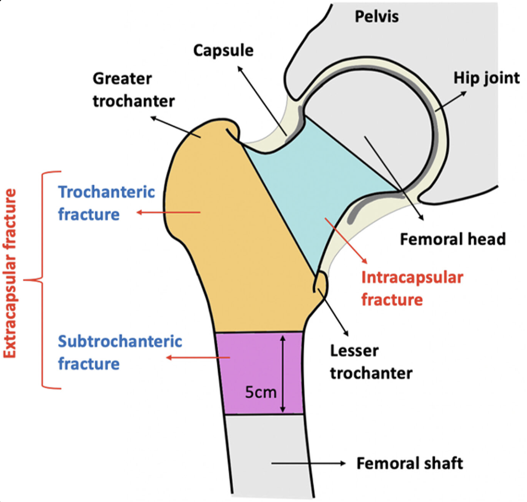
Anatomy of the proximal femur and the main types of hip fracture. Intracapsular fractures (blue) occur within the hip joint capsule at the femoral neck. Extracapsular fractures sit outside the capsule and include trochanteric fractures (orange), between the greater and lesser trochanter, and subtrochanteric fractures (purple), within about 5 cm below the lesser trochanter.
WHY ARE WOMEN AT GREATER RISK?
Most hip fractures in older adults happen after a low-energy fall from standing height, usually landing sideways onto the hip [1,3]. Whether that fall results in fracture depends largely on the strength of the bone underneath. With age, bone mass gradually declines in both sexes. However, osteoporosis, a condition in which bones become thin, porous, and brittle, is considerably more common in women [3]. Furthermore, hormonal shifts of menopause accelerate bone loss, underpinning the steep increase in women's fracture risk in later life [1,3].
In addition to gradual bone loss, women face higher fall rates than men across age groups, and research suggests that differences in balance control, faster loss of muscle mass after menopause, more sedentary lifestyles, and poorer nutrition may contribute to this [1]. Older women are also more likely than men to experience depression and use prescribed psychiatric medications, both of which are recognized risk factors for falls [1]. Combined with women's longer life expectancy, these factors increase cumulative exposure to fall-related risks, making women more vulnerable to serious consequences from a single fall as they age [1].
The types of hip fractures women sustain also change with age. Hip fractures are generally divided into two categories: 1) intracapsular fractures, which occur within the hip joint capsule at the femoral neck, and 2) extracapsular fractures, which occur outside the capsule in the trochanteric region of the upper femur [1]. Intertrochanteric fractures, a type of extracapsular fracture that occurs between the greater and lesser trochanters, are particularly associated with osteoporosis [1]. Research has found that the proportion of women experiencing this fracture type increased from approximately 52% among those aged 65-74 to nearly 59% among those over 75, while the proportion in men remains relatively stable [1]. This suggests that the difference may reflect sex-related differences in the extent and progression of bone loss, as well as fall frequency [1].
WHAT HAPPENS AFTER A HIP FRACTURE?
Most hip fractures in older adults require surgery to relieve pain and restore mobility [3]. While surgery is often the best option, it also exposes patients to a range of complications, including increased risk of infection, blood clots in the legs or lungs, pressure sores, urinary tract infections, and pneumonia [3]. Ultimately outcomes of surgery are shaped by a patient's age and sex, as well as by the health conditions they were already living with, for instance heart or lung disease, diabetes, cognitive decline, or frailty [3].
Beyond risk of surgical complications, the risk of death after a hip fracture is substantial. A population-based Finnish study following 428 hip fracture patients aged 65 and older, roughly three-quarters of whom were women, found that about 1 in 10 had died within 30 days of surgery and more than 1 in 4 within the first year [4]. Over the full follow-up period, hip fracture patients died at about three times the rate of the age and sex matched general population, and this excess risk appeared across every major cause of death, with circulatory disease and dementia emerging as the most common [4].
Notably, this study found that men faced a higher risk of death after hip fracture than women, however this does not necessarily mean that hip fractures are less serious for women. Because women have much higher hip fracture incidence rates overall, they account for the majority of people living with, and dying after, this injury [1,4]. Furthermore, women in this study were older at the time of fracture and were about twice as likely as men to already be living with dementia, rendering women more likely to enter recovery from an already vulnerable position [4].
For those who survive, the effects of hip fracture are often lasting. Recovery in older or frail adults can take months, and prolonged inactivity during that time can lead to further muscle atrophy, which in turn decreases stability and balance, thereby increasing the risk of a subsequent fall [3]. Many older adults never fully regain their level of activity and independence [3].
WHAT DOES RECOVERY LOOK LIKE?
Physical therapy typically starts almost immediately after surgery, not only to help patients regain mobility but also to reduce the complications associated with being immobile, such as infection and blood clots [3]. Adequate nutrition, particularly sufficient protein intake to support muscle mass maintenance, supports healing, while occupational therapy ensures that patients stay mentally and emotionally engaged in order to lower the risk of depression during a long recovery period [3].
Pain management is another crucial, and often overlooked, part of recovery in older adults [3]. Clear communication about pain, often involving describing it on a scale from 1 to 10, allows healthcare teams to respond appropriately and supports better participation in rehabilitation [3].
The structure of care after surgery also matters. There is evidence that active, team-based geriatric rehabilitation can shorten hospital stays and improve recovery of daily activities, indicating a need for stronger coordination between hospital and community care programs after discharge [4]. For families and caregivers, this often means planning ahead for the additional support a patient may need following their hospital stay [3].
HOW IS THE RELEVANT IN WOMEN'S HEALTHCARE?
Given the far-reaching consequences of hip fractures, identifying women at increased risk before a fall occurs is an important goal of preventive care. Bone mineral density screening using dual-energy X-ray absorptiometry (DEXA) scans is recommended for women beginning at age 65, or earlier in the presence of risk factors such as low body weight, smoking, or long-term corticosteroid use [2]. However, although DEXA scans help identify osteoporosis and low bone density, bone density is only one of several factors that contribute to hip fracture risk [2].
Simple clinical tools and scales can help fill this gap. One example, the FRAMO (Fracture and Mortality) Index, was validated in nearly 1,500 older Swedish women and uses just four questions: whether a woman is 80 or older, whether she weighs less than about 60 kg (132 lb), whether she can stand up from a chair five times without using her arms, and whether she has had a fracture after age 40 [2]. Clinical tools such as these are relatively easy to implement in primary care and can help identify women who may benefit from closer monitoring and preventive interventions [2].
Once a woman is identified as high risk, several practical steps may lower her chances of a fracture. These include exercise and balance programs, stopping smoking and reducing alcohol use, and making changes to the home environment to reduce fall hazards, such as installing grab bars in showers and improving awareness of slippery surfaces [2]. Staying physically active in midlife has also been linked to a lower risk of hip fracture later on, likely through increased maintenance of muscle strength, coordination, and balance [1].
Ultimately, from a women's health perspective, these findings highlight the need to look beyond bone density alone. Menopause-related bone loss, fall risk, mental health, medication use, cognitive health, and social support all intersect to shape a woman's vulnerability to hip fracture and her recovery trajectory [1, 3, 4].
REFERENCES
Alpantaki K, Papadaki C, Raptis K, Dretakis K, Samonis G, Koutserimpas C. Gender and age differences in hip fracture types among elderly: a retrospective cohort study. Maedica (Bucur). 2020;15(2):185-190. doi:10.26574/maedica.2020.15.2.185. https://pmc.ncbi.nlm.nih.gov/articles/PMC7482680/
Ebell MH. Predicting hip fracture risk in older women. Am Fam Physician. 2007;76(2):273-275. Accessed November 2, 2026. https://www.aafp.org/afp/2007/0715/p273
Ko E, Glazier E. Hip fractures in elderly often lead to bad outcomes. UCLA Health. Published November 23, 2020. Accessed November 2, 2026. https://www.uclahealth.org/news/article/hip-fractures-in-elderly-often-lead-to-bad-outcomes
Panula J, Pihlajamäki H, Mattila VM, et al. Mortality and cause of death in hip fracture patients aged 65 or older: a population-based study. BMC Musculoskelet Disord. 2011;12:105. doi:10.1186/1471-2474-12-105. https://pmc.ncbi.nlm.nih.gov/articles/PMC3118151/
